Translational Quantitative Imaging Center
Our work
Advanced Translational Imaging Research Core
The Sports Medicine group at UCSF utilizes advanced biomedical imaging techniques to study different conditions of the knee, shoulder, and hip. Magnetic resonance imaging (MRI) scans produce high-resolution three-dimensional images. Specialized MRI sequences can also provide detailed information about the biochemical composition of tissue, tissue architecture, or the function of joints. Our Sports Medicine group closely collaborates with the UCSF Department of Radiology and the MQIR (Musculoskeletal Quantitative Imaging Research) group to leverage these advanced technologies to better evaluate patients and the effects of non-surgical and surgical treatment.
Current projects
Knee Imaging
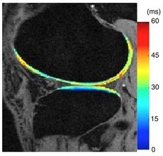
Knee ligament injuries and cartilage injuries are common conditions, especially in active people. Following anterior cruciate ligament (ACL) tears, patients are at an increased risk of developing knee arthritis. We can track the composition of cartilage using two advanced MRI sequences: T1rho and T2 mapping.10 The T1rho mapping sequence can measure the content of proteoglycans, which are an important component of healthy cartilage. The T2 mapping sequences gives information on the structure of collagen in cartilage. Both sequences can detect breakdown of cartilage early in the degenerative process (Figure 1). We have used these sequences to monitor improvement after cartilage repair surgery and evaluate for early cartilage breakdown in patients with ACL tears.1, 12, 13
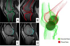
We have also used kinematic MRI to evaluate the alignment and motion of the knee after injury to the ACL and following ACL reconstruction surgery.5 We obtain MRI scans with a weight applied to the foot, to simulate standing in the scanner.3 We obtain images with the knee straight and then also bent. We can then reconstruct three-dimensional models of the knee and better understand the complex function of the knee through motion (Figure 2).6, 8 We have linked abnormal knee motion to early cartilage breakdown by combining these imaging technologies.6, 15
Shoulder Imaging
Our research has applied advanced imaging techniques to study patients with rotator cuff injuries. The muscles of the rotator cuff undergo degenerative changes following rotator cuff tears, with the muscle both shrinking in size (atrophy) and being replaced by fat (fatty infiltration).2, 4 Both degenerative changes are associated with worse outcomes after surgical treatment. We have used an advanced MRI sequence, IDEAL, to measure the fat content in the shoulder muscles.11 We have shown that increasing fat content is seen in larger rotator cuff tears.9 We have also studied how the fat content changes after surgical repair of a rotator cuff tear and demonstrated that lower fat content prior to surgical repair is associated with a higher chance of successful tendon repair.7
Hip Imaging
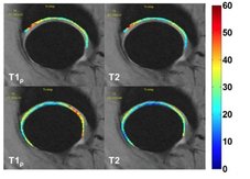
We have applied the T1rho and T2 mapping sequences to track the cartilage health of the hip. In femoroacetabular impingement (FAI), bony mismatch between the femoral head (ball) and acetabulum (socket) are associated with labral tears and cartilage breakdown. We have shown that T1rho and T2 mapping can identify hip cartilage injuries better than traditional MRI (Figure 3).14
Publications
- Amano K, Pedoia V, Su F, Souza RB, Li X, Ma CB. Persistent Biomechanical Alterations After ACL Reconstruction Are Associated With Early Cartilage Matrix Changes Detected by Quantitative MR. Orthopaedic Journal of Sports Medicine. 2016;4(4).
- Barry JJ, Lansdown DA, Cheung S, Feeley BT, Ma CB. The relationship between tear severity, fatty infiltration, and muscle atrophy in the supraspinatus. Journal of shoulder and elbow surgery. 2013;22(1):18-25.
- Carpenter RD, Majumdar S, Ma CB. Magnetic Resonance Imaging of 3-Dimensional In Vivo Tibiofemoral Kinematics in Anterior Cruciate Ligament–Reconstructed Knees. Arthroscopy: The Journal of Arthroscopic & Related Surgery. 2009;25(7):760-766.
- Cheung S, Dillon E, Tham S-C, et al. The Presence of Fatty Infiltration in the Infraspinatus: Its Relation With the Condition of the Supraspinatus Tendon. Arthroscopy: The Journal of Arthroscopic & Related Surgery. 2011;27(4):463-470.
- Haughom B, Schairer W, Souza RB, Carpenter D, Ma CB, Li X. Abnormal tibiofemoral kinematics following ACL reconstruction are associated with early cartilage matrix degeneration measured by MRI T1rho. The Knee. 2012;19(4):482-487.
- Lansdown DA, Allen C, Zaid M, et al. A comprehensive in vivo kinematic, quantitative MRI and functional evaluation following ACL reconstruction — A comparison between mini-two incision and anteromedial portal femoral tunnel drilling. The Knee. 2015;22(6):547-553.
- Lansdown DA, Lee S, Sam C, Krug R, Feeley BT, Ma CB. A Prospective, Quantitative Evaluation of Fatty Infiltration Before and After Rotator Cuff Repair. Orthopaedic journal of sports medicine. 2017;5(7):2325967117718537.
- Lansdown DA, Zaid M, Pedoia V, et al. Reproducibility measurements of three methods for calculating in vivo MR-based knee kinematics. Journal of Magnetic Resonance Imaging. 2015;42(2):533-538.
- Lee S, Lucas RM, Lansdown DA, et al. Magnetic resonance rotator cuff fat fraction and its relationship with tendon tear severity and subject characteristics. Journal of Shoulder and Elbow Surgery. 2015;24(9):1442-1451.
- Li X, Pedoia V, Kumar D, et al. Cartilage T 1ρ and T 2 relaxation times: longitudinal reproducibility and variations using different coils, MR systems and sites. Osteoarthritis and cartilage. 2015;23(12):2214-2223.
- Nardo L, Karampinos DC, Lansdown DA, et al. Quantitative assessment of fat infiltration in the rotator cuff muscles using water-fat MRI. Journal of Magnetic Resonance Imaging. 2014;39(5):1178-1185.
- Pedoia V, Su F, Amano K, et al. Analysis of the articular cartilage T1ρ and T2 relaxation times changes after ACL reconstruction in injured and contralateral knees and relationships with bone shape. Journal of Orthopaedic Research. 2016.
- Theologis AA, Schairer WW, Carballido-Gamio J, Majumdar S, Li X, Ma CB. Longitudinal analysis of T1p and T2 quantitative MRI of knee cartilage laminar organization following microfracture surgery. The Knee. 2012;19(5):652-657.
- Wyatt C, Kumar D, Subburaj K, et al. Cartilage T1ρ and T2 Relaxation Times in Patients With Mild‐to‐Moderate Radiographic Hip Osteoarthritis. Arthritis & Rheumatology. 2015;67(6):1548-1556.
- Zaid M, Lansdown D, Su F, et al. Abnormal tibial position is correlated to early degenerative changes one year following ACL reconstruction. Journal of Orthopaedic Research. 2015;33(7):1079-1086.